You've been living with lower back pain for months, maybe even years. You've seen multiple doctors, tried physical therapy, and perhaps even undergone imaging studies. Yet the pain persists, radiating through your lower back and into your buttocks and legs. Sound familiar? You may be among the countless individuals whose sacroiliac joint dysfunction has gone undiagnosed—a condition responsible for up to 25% of chronic lower back pain cases.
The sacroiliac joint is one of medicine's great mimics, producing symptoms that can easily be mistaken for disc herniation, sciatica, or general back strain. This diagnostic challenge leaves many patients frustrated, undertreated, and wondering if they'll ever find relief. Understanding this often-overlooked condition is the first step toward effective treatment and recovery.
What Is the Sacroiliac Joint?
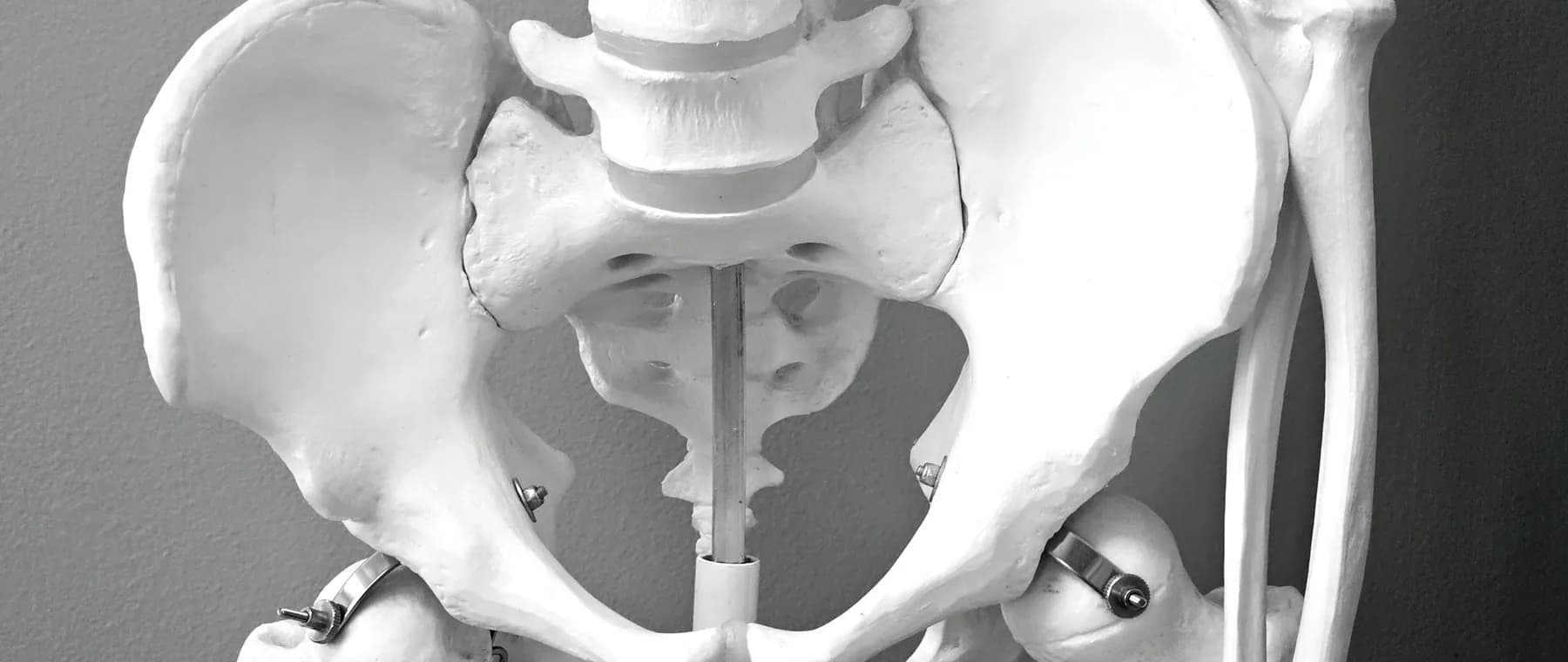
The sacroiliac joints are two small but mighty structures located where your spine meets your pelvis. These joints connect the sacrum (the triangular bone at the base of your spine) to the iliac bones of your pelvis on either side. Despite their relatively small range of motion, sacroiliac joints play a crucial role in transferring weight and forces between your upper body and legs.
Unlike many joints in your body, the SI joints are designed more for stability than mobility. They're surrounded by some of the strongest ligaments in the body and are reinforced by numerous muscles. When functioning properly, these joints allow just enough movement to absorb shock during walking and other activities while maintaining pelvic stability.
Problems arise when these joints become either too mobile (hypermobile) or too restricted (hypomobile). Inflammation, injury, arthritis, or biomechanical stress can all disrupt normal SI joint function, leading to a condition known as sacroiliac joint dysfunction or SI joint pain.
Why SI Joint Pain Is Frequently Misdiagnosed
The diagnostic challenge with SI joint dysfunction stems from its symptom overlap with more commonly recognized conditions. The pain typically manifests in the lower back, buttocks, and can radiate down one or both legs—a pattern nearly identical to sciatica caused by disc herniation. This similarity often leads practitioners to focus on the spine while overlooking the SI joint as a potential pain generator.
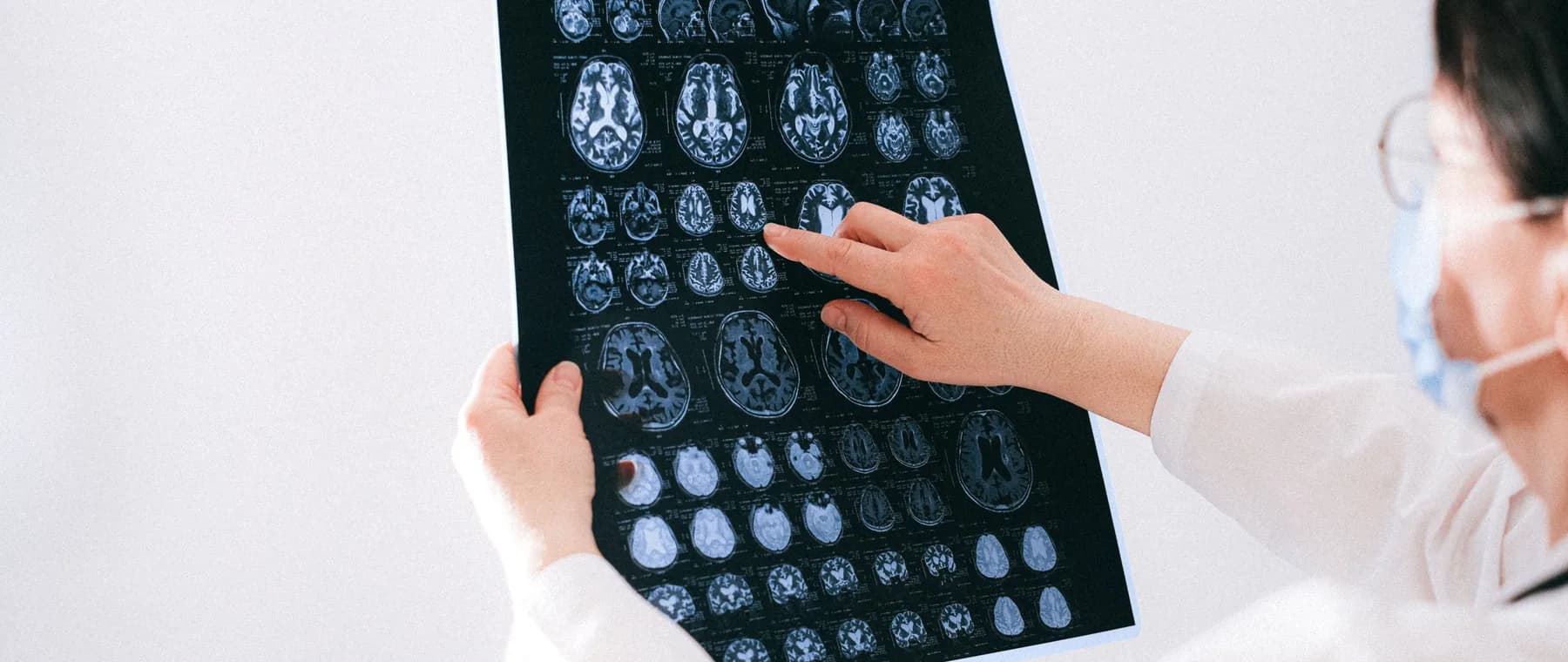
Standard imaging studies add another layer of complexity. X-rays and MRI scans excel at revealing disc problems and spinal abnormalities, but they rarely show SI joint dysfunction unless significant arthritis is present. Many patients receive diagnoses based on incidental findings on imaging that may not be the true source of their pain, while the actual culprit—the SI joint—remains unexamined.
The location of SI joint pain can also be misleading. While some patients experience localized tenderness directly over the joint, others feel pain referred to distant areas including the groin, hip, or even down to the ankle. This wide distribution of symptoms can send clinicians and patients alike on a diagnostic wild goose chase.
"Up to 25% of chronic lower back pain cases can be attributed to sacroiliac joint dysfunction, yet it remains one of the most frequently overlooked diagnoses in spine care."
Recognizing the Signs and Symptoms
While SI joint pain can be subtle and variable, certain patterns emerge that can help identify this condition. Understanding these characteristic features is essential for both patients and healthcare providers.
The hallmark of SI joint dysfunction is pain localized to the lower back and buttock, usually on one side. Patients often describe being able to point with one finger to the exact spot that hurts, typically just below and to the side of the lower back. This pain frequently worsens with specific movements and positions:
Additionally, many patients report that their symptoms feel different from typical back pain—often describing sensations of instability, as if their pelvis might "give out," or a deep, aching quality that differs from sharp nerve pain associated with disc problems.
Risk Factors and Contributing Conditions
Certain factors increase the likelihood of developing SI joint dysfunction. Women are more susceptible than men, partly due to hormonal influences and structural differences in pelvic anatomy. Pregnancy, in particular, significantly increases risk as hormones cause ligament laxity and the growing uterus shifts the body's center of gravity, placing additional stress on the SI joints.
Previous lumbar spine surgery can also predispose individuals to SI joint problems. When spinal fusion eliminates motion at certain vertebral levels, compensatory stress is transferred to adjacent structures, including the SI joints. Studies suggest that up to 40% of patients who continue experiencing pain after lumbar fusion may have developed SI joint dysfunction.
Other risk factors include leg length discrepancies, inflammatory conditions like ankylosing spondylitis, traumatic injuries such as falls or motor vehicle accidents, and repetitive stress from activities involving twisting or uneven loading of the pelvis.
Athletes who participate in running, golf, or other sports involving rotational movements may be particularly vulnerable. The cumulative effect of repetitive motion, combined with any of these predisposing factors, can gradually compromise SI joint function over time.
Accurate Diagnosis: The Key to Effective Treatment
Properly diagnosing SI joint dysfunction requires a thorough clinical evaluation that goes beyond standard imaging. The diagnostic process typically begins with a detailed history and physical examination, including specific provocative tests designed to stress the SI joint and reproduce symptoms.
These physical examination maneuvers include tests like the FABER (flexion, abduction, external rotation), Gaenslen's, thigh thrust, and compression tests. When three or more of these tests are positive, the likelihood of SI joint involvement increases significantly. However, because no single test is definitive, clinicians often rely on a combination of findings.
The diagnostic gold standard remains a fluoroscopically-guided SI joint injection using local anesthetic. If a patient experiences significant pain relief following this injection (typically 75% or greater reduction), it confirms the SI joint as the primary pain generator. This diagnostic injection not only validates the diagnosis but can also provide therapeutic benefit.
It's worth noting that SI joint dysfunction often coexists with other spinal conditions. A comprehensive evaluation should consider all potential pain sources to develop the most effective treatment strategy. In Miami, FL, specialized pain management clinics utilize these advanced diagnostic techniques to accurately identify SI joint problems and distinguish them from other causes of lower back pain.
Treatment Options and Path Forward
Once properly diagnosed, SI joint dysfunction can be effectively managed through a variety of treatment approaches. Conservative management typically begins with physical therapy focused on strengthening the muscles that support the pelvis and improving joint mechanics. Manual therapy techniques and exercises to address muscle imbalances often provide significant relief.
When conservative measures prove insufficient, interventional treatments offer additional options. SI joint injections with corticosteroids can reduce inflammation and provide extended pain relief. Radiofrequency ablation, which uses heat to interrupt pain signals from the joint, may offer longer-lasting results for appropriate candidates.
For severe, chronic cases that don't respond to other interventions, minimally invasive SI joint fusion procedures have shown promising outcomes. These surgical options stabilize the joint while preserving as much function as possible.
The key to successful treatment lies in accurate diagnosis and a personalized approach that addresses each patient's specific presentation and goals. With proper identification and management, most individuals with SI joint dysfunction can experience substantial improvement and return to their normal activities.
If you've been struggling with persistent lower back pain that hasn't responded to conventional treatments, sacroiliac joint dysfunction may be the missing piece of your diagnostic puzzle. Don't let another year pass living with unnecessary pain. Dr. Steinberger uses image-guided diagnostic blocks to establish whether the SI joint is the source of your pain before building a treatment plan around that answer. Your path to relief may be closer than you think—it starts with the right diagnosis.