
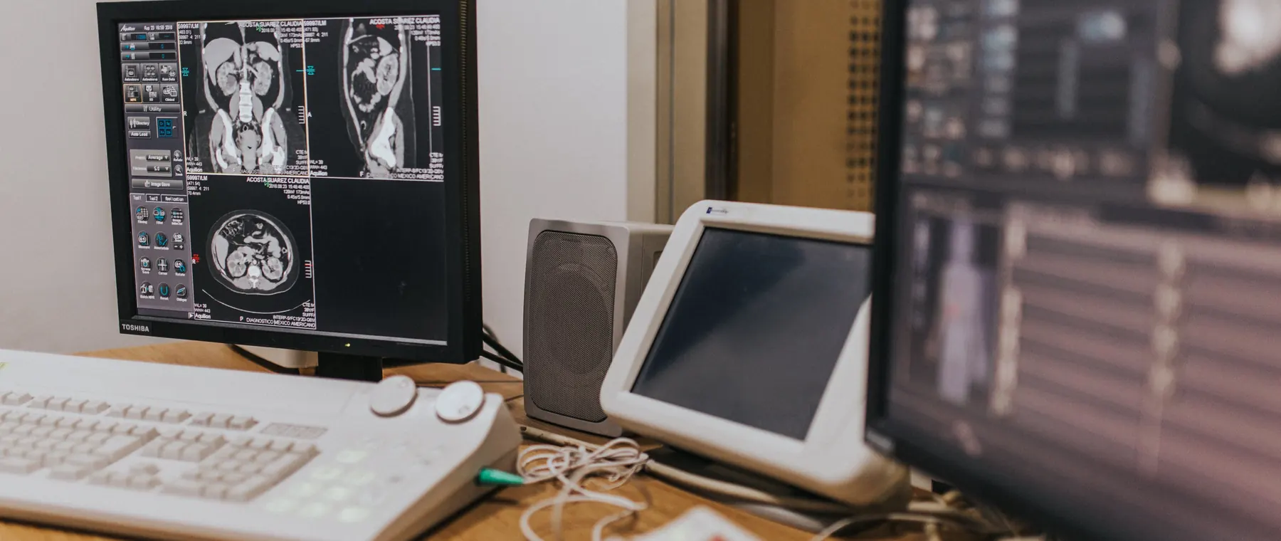
When chronic pain disrupts your daily life, finding the source can feel like solving a complex puzzle. You know something is wrong—your body is sending urgent signals—but understanding exactly what's causing your discomfort often requires looking beneath the surface. This is where medical imaging becomes an invaluable tool, offering healthcare providers a window into the body's internal structures to identify the root causes of pain and develop targeted treatment strategies.
Yet for many patients, the world of medical imaging can seem overwhelming. X-rays, MRIs, CT scans, ultrasounds—each technology serves a unique purpose in the diagnostic process. Understanding how these tools work and what they reveal can empower you to be an active participant in your care journey, asking informed questions and making confident decisions about your treatment path.
Why Imaging Matters in Pain Diagnosis
Pain is a complex experience that results from intricate interactions between nerves, tissues, and the brain. While a thorough physical examination and detailed medical history provide essential information, they cannot always reveal structural abnormalities, inflammation, or degenerative changes that may be contributing to your symptoms.
Medical imaging bridges this gap by providing objective visual evidence of what's happening inside your body. Whether it's a herniated disc pressing on a nerve, arthritis eroding joint surfaces, or soft tissue inflammation, imaging technologies allow physicians to see conditions that would otherwise remain hidden. This visibility is crucial not only for accurate diagnosis but also for monitoring disease progression and evaluating treatment effectiveness over time.
In Miami, FL, where residents lead active lifestyles—from water sports to urban recreation—imaging plays a particularly important role in identifying sports-related injuries, degenerative conditions accelerated by humidity and activity levels, and age-related changes that manifest as chronic pain.
Common Imaging Technologies and What They Reveal
Different imaging modalities excel at visualizing different aspects of anatomy and pathology. Understanding the strengths of each helps explain why your physician might order one test over another.
X-rays (Radiography)
X-rays remain the most common first-line imaging tool due to their accessibility, speed, and cost-effectiveness. They excel at showing bone structures, making them ideal for detecting fractures, arthritis, bone spurs, and alignment issues. However, X-rays provide limited information about soft tissues like muscles, ligaments, and nerves.
Magnetic Resonance Imaging (MRI)
MRI uses powerful magnets and radio waves to create detailed images of both hard and soft tissues. It's the gold standard for visualizing herniated discs, spinal cord compression, ligament tears, cartilage damage, and soft tissue masses. MRI is particularly valuable in diagnosing conditions affecting the spine, joints, and complex anatomical regions where multiple structures interact.
Computed Tomography (CT Scans)
CT scans use X-ray technology combined with computer processing to create cross-sectional images of the body. They're excellent for evaluating complex bone fractures, spinal stenosis, and conditions requiring detailed anatomical precision. CT imaging is faster than MRI and can be performed on patients with metal implants or pacemakers.
Ultrasound
Ultrasound uses sound waves to create real-time images of soft tissues, blood flow, and fluid collections. In pain management, ultrasound is increasingly used for guiding injections with precision, evaluating joint inflammation, and assessing tendon and muscle injuries. It's radiation-free and can be performed at the point of care.
"Imaging doesn't just show us what's wrong—it guides us toward what's right, helping tailor treatment plans that address the specific source of your pain rather than just masking symptoms."
When Is Imaging Necessary?
Not every instance of pain requires immediate imaging. In fact, many acute pain episodes—such as minor strains or muscle tension—resolve with conservative treatment and time. However, certain "red flags" warrant prompt imaging evaluation:
- Pain following significant trauma or injury
- Persistent pain lasting beyond 4-6 weeks despite conservative treatment
- Pain accompanied by neurological symptoms like numbness, weakness, or loss of bowel/bladder control
- Suspicion of infection, tumor, or other serious underlying condition
- Progressive worsening of symptoms over time
- Pain that significantly impacts quality of life and daily function
Your physician will consider your complete clinical picture—symptoms, medical history, physical examination findings, and risk factors—when determining whether imaging is appropriate and which modality would be most informative.
Understanding Your Imaging Results
Receiving imaging results can be both relieving and anxiety-provoking. It's important to remember that imaging findings must always be interpreted in the context of your symptoms and clinical presentation.
Studies have shown that many people without any pain have imaging abnormalities—bulging discs, arthritis, and degenerative changes are common findings in asymptomatic individuals, especially as we age. Conversely, some people experience significant pain despite relatively normal imaging. This is why the conversation with your healthcare provider about what the images mean for your specific situation is so crucial.
Your provider will explain which findings are likely contributing to your pain, which are incidental, and how the imaging results inform your treatment plan. Don't hesitate to ask questions: What exactly does this finding mean? Is it the cause of my pain? What are my treatment options? How will we know if treatment is working?
How Imaging Guides Treatment Decisions
Beyond diagnosis, imaging plays a vital role in treatment planning and execution. Interventional pain procedures—such as epidural steroid injections, nerve blocks, and radiofrequency ablation—often rely on image guidance to ensure medication or treatment is delivered precisely to the target area.
Fluoroscopy (real-time X-ray), ultrasound, and CT guidance allow physicians to navigate complex anatomy safely and effectively, maximizing therapeutic benefit while minimizing risks. This precision medicine approach represents a significant advancement over older "blind" injection techniques.
Additionally, repeat imaging may be ordered to monitor disease progression, assess healing after injury, or evaluate the structural effects of treatment over time. These follow-up studies provide objective measures of improvement or indicate when treatment strategies need adjustment.
Taking the Next Step
If you're living with chronic or persistent pain, understanding the role of imaging in diagnosis and treatment can help you feel more confident and informed as you seek care. Remember that imaging is just one piece of the puzzle—it works alongside your symptom description, physical examination, and medical history to create a complete picture.
At Gables Pain in Miami, FL, Dr. Steinberger uses imaging findings as part of a comprehensive approach to pain diagnosis and management. We take the time to explain what your images show, how findings relate to your symptoms, and what treatment options are available based on your unique situation.
You don't have to navigate pain alone. Whether you're seeking a diagnosis for new symptoms or looking for more effective management of ongoing pain, Dr. Steinberger is here to provide expert, compassionate care tailored to your individual needs.